Definition
Abdominal ultrasound is an imaging procedure used to examine the internal organs of the abdomen, including the liver, gallbladder, spleen, pancreas, and kidneys. The blood vessels that lead to some of these organs can also be looked at with ultrasound.

How the test is performed
An ultrasound machine sends out high-frequency sound waves, which reflect off body structures to create a picture. A computer receives these reflected waves and uses them to create an image. Unlike x-rays or CT scans, there is no ionising radiation exposure with this test.
You will be lying down for the procedure. A clear, water-based conducting gel is applied to the skin over the abdomen. This helps with the transmission of the sound waves. A handheld probe called a transducer is then moved over the abdomen.
You may be asked to change position so that the sonographer can examine different areas. You may also be asked to hold your breath for short periods of time during the examination.
The procedure usually takes less than 30 minutes.
How to prepare for the test
Preparation for the procedure depends on the nature of the problem and your age. Usually patients are asked to not eat or drink for several hours before the examination. The receptionist you make your booking with will advise you about specific preparation.
How the test will feel
There is little discomfort. The conducting gel may feel slightly cold and wet.
Why the test is performed
Your doctor may order this test to:
- Determine the cause of abdominal pain
- Learn why there is swelling of an abdominal organ
- Look for stones in the gallbladder or kidney
- The specific reason for the test will depend on your symptoms.
Normal Values
The organs examined are normal in appearance.
What abnormal results mean
Some conditions identified on ultrasound include:
- Abdominal aortic aneurysm (blood vessel becomes abnormally large or balloon outward)
- Cholecystitis (inflammation of the gallbladder)
- Gallstones (crystalline concretion within the gallbladder)
- Hydronephrosis (obstruction of the free flow of urine from the kidney)
- Kidney stones (crystalline concretion within the kidney)Spleen enlargement (splenomegaly) Pancreatitis (inflammation of the pancreas)
What the risks are
There is no documented risk. No ionising radiation exposure is involved.
From the moment you request an appointment to the final referral, we aim to make your experience as smooth and comfortable as possible. Here is our working process for all DiagnostiCare patients.
-

Request Appointment
-
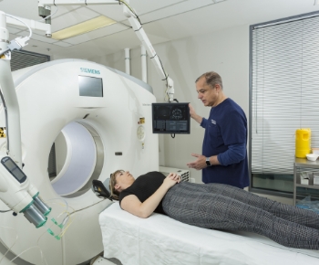
Patient Examination
-
Results To Referrer


-
Use of low-radiation techniques for safety.
-
Capable of performing complex imaging procedures.
-
Bulk-billing available for eligible patients.
-
Strong commitment to patient privacy and confidentiality.
-
Offering both walk-in and appointment-based services.
-
Equipped with the latest 3D imaging technology.
-
Suite 46 Level 1 Milleara Mall,
235 Milleara Road East Keilor VIC 3033 -
Monday - Friday 8.00am - 6.00pm Saturday 8.00am - 1.00pm

